The practical examination
Weave Viva
Prepare in depth.
Return to the essentials.
The full practical book and the hundred-map companion. Original text or illustrated edition, together in one place.
For MD and DNB psychiatry revision.
The depth for a full preparation.
Nine parts, from case presentation and neurological examination to OSCEs, viva tables and spotters. Choose the original text or the illustrated practical book.
Look inside the deep book
Real pages from the original and illustrated editions. Select an image to enlarge it. Image source and licence links accompany the illustrated sample.
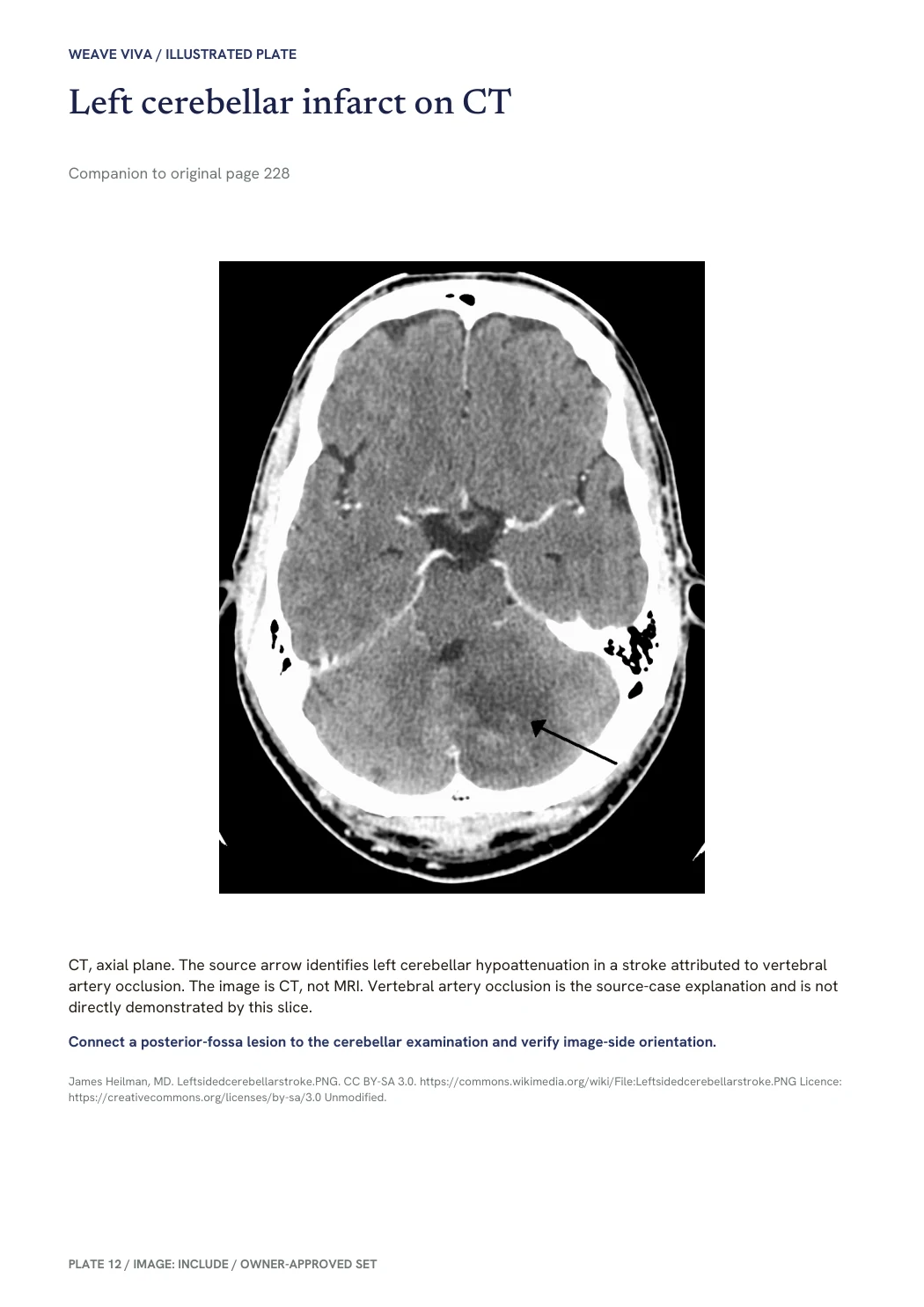
CT, axial plane. The source arrow identifies left cerebellar hypoattenuation in a stroke attributed to vertebral artery occlusion. The image is CT, not MRI. Vertebral artery occlusion is the source-case explanation and is not directly demonstrated by this slice.
James Heilman, MD. Leftsidedcerebellarstroke.PNG. CC BY-SA 3.0. https://commons.wikimedia.org/wiki/File:Leftsidedcerebellarstroke.PNG Licence: https://creativecommons.org/licenses/by-sa/3.0 Unmodified. Image source · CC BY-SA 3.0

An unmodified page from the original edition.
Weave. Original book page; credits and rights are retained in the PDF.
The hundred for a focused revision.
One hundred answer maps in a compact practical companion. Choose the original maps or the edition with selected illustrations alongside them.
Look inside the concise companion
Real pages from the original and illustrated editions. Select an image to enlarge it. Image source and licence links accompany the illustrated sample.
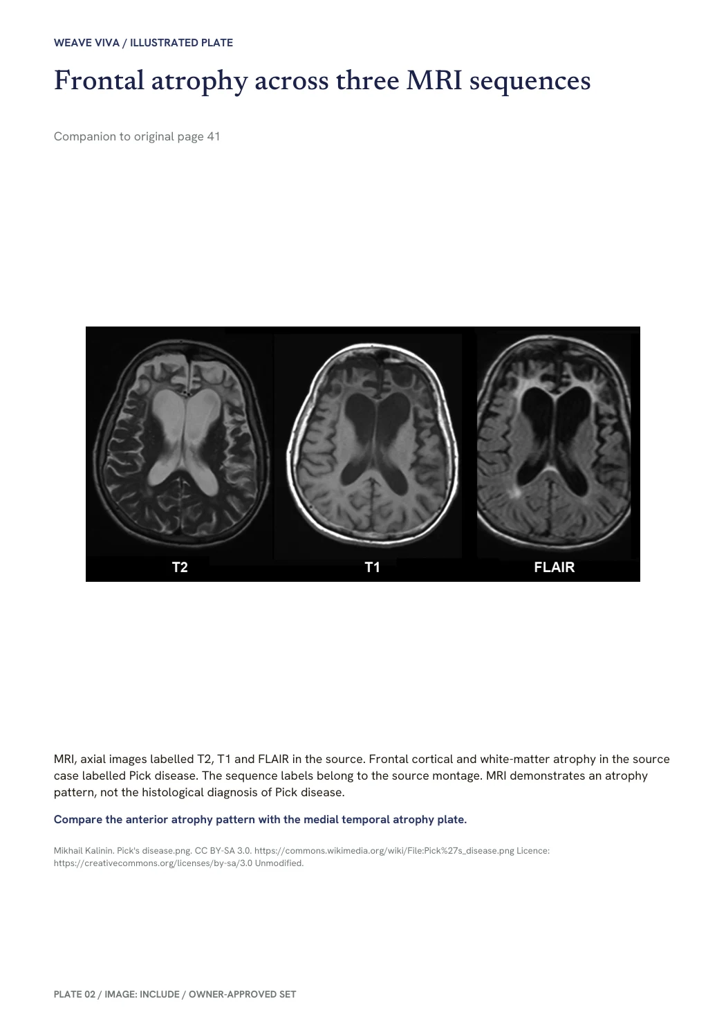
MRI, axial images labelled T2, T1 and FLAIR in the source. Frontal cortical and white-matter atrophy in the source case labelled Pick disease. The sequence labels belong to the source montage. MRI demonstrates an atrophy pattern, not the histological diagnosis of Pick disease.
Mikhail Kalinin. Pick's disease.png. CC BY-SA 3.0. https://commons.wikimedia.org/wiki/File:Pick%27s_disease.png Licence: https://creativecommons.org/licenses/by-sa/3.0 Unmodified. Image source · CC BY-SA 3.0

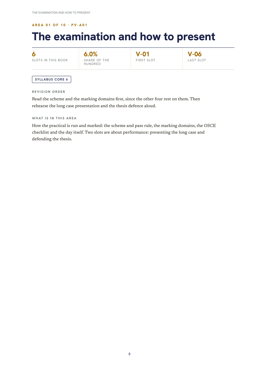
An unmodified page from the original edition.
Weave. Original book page; credits and rights are retained in the PDF.
Use the book that fits the reading.
The deep book develops the practical topics in detail. The concise companion is for returning to the answer maps. Illustrated editions add selected plates; they do not claim complete image or spotter coverage.
These are educational revision companions. Check current clinical guidance, local examination requirements and the source references. Captions, image credits and licence statements stay with each book.
Explore Weave 100 and Weave 100+ for theory